In modern ophthalmology, precision is everything. When surgeons plan cataract procedures or evaluate complex eye conditions, even a fraction of a millimeter can influence visual outcomes. That is where DGH A technology plays a critical role. Known in clinical settings as the Scanmate A, this diagnostic device has earned a solid reputation among U.S. eye care professionals for reliability, portability, and measurement accuracy.
Manufactured by DGH Technology Inc, the Scanmate A is an A-Scan ultrasound biometer designed specifically for ophthalmic use. While optical biometers have become more common in larger surgical centers, ultrasound A-Scan devices remain indispensable in many practices across the United States.
What Is DGH A?
At its core, DGH A refers to an ophthalmic A-Scan ultrasound device used to measure internal eye structures. These measurements are essential for calculating intraocular lens (IOL) power prior to cataract surgery.
The system operates by sending high-frequency sound waves into the eye. When these sound waves encounter tissue boundaries, they reflect back to the probe. The device calculates the time it takes for those echoes to return and converts that information into precise distance measurements.
Unlike general medical ultrasound machines, this device is designed exclusively for eye diagnostics. That specialization contributes to its measurement accuracy and clinical reliability.
Why Accurate Eye Measurements Matter
Before cataract surgery, surgeons must determine the exact power of the artificial lens that will replace the clouded natural lens. A small error in axial length measurement — even 0.1 mm — can significantly affect postoperative vision.
Accurate measurements directly influence:
- Post-surgery visual clarity
- Reduced need for corrective eyewear
- Patient satisfaction
- Fewer refractive surprises
Because cataract surgery is one of the most commonly performed procedures in the United States, dependable biometric tools are essential in both private practices and hospital settings.
How the Technology Works
Understanding A-Scan Ultrasound
A-Scan stands for “amplitude scan.” The ultrasound probe emits a narrow beam of sound energy. As the sound waves travel through the eye, they encounter different tissues such as:
- Cornea
- Aqueous humor
- Lens
- Vitreous body
- Retina
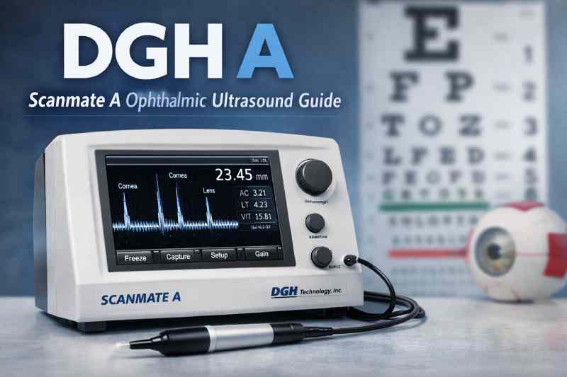
Each tissue reflects sound differently. The system records these reflections and displays them as spikes on a graph. The distance between these spikes represents anatomical measurements.
Measurement Parameters
The device typically measures:
- Axial length
- Anterior chamber depth
- Lens thickness
- Vitreous length
These values are entered into IOL calculation formulas to determine the correct lens implant power.
Contact vs. Immersion Mode
Clinicians can choose between two techniques:
Contact Mode
The probe gently touches the cornea. This method is quick and commonly used in everyday practice.
Immersion Mode
A fluid interface prevents direct corneal compression, increasing accuracy. This method is preferred in more sensitive or high-precision cases.
Many U.S. clinics use immersion for surgical planning and contact mode for routine measurements.
Clinical Applications
The most common use of dgh a technology is preoperative cataract assessment. However, its applications extend beyond that single procedure.
Cataract Surgery Planning
The primary purpose is IOL power calculation. Reliable axial length measurement is critical to achieving target refraction after surgery.
Dense Cataract Cases
Optical biometers often struggle with advanced or dense cataracts because light cannot pass clearly through the cloudy lens. Ultrasound, however, is not affected by lens opacity. This makes the system particularly valuable when optical measurement fails.
Pediatric Ophthalmology
Children may have difficulty maintaining fixation for optical devices. Ultrasound A-Scan provides a practical alternative.
Research and Clinical Studies
Because of its repeatable measurement capability, it is often used in academic environments for longitudinal eye studies.
Why It Remains Relevant in Modern Clinics
Despite the growth of optical coherence biometry, dgh a devices remain widely used in the United States. Several factors explain this continued demand.
Reliability in Challenging Cases
Ultrasound works even when media opacity blocks optical signals. This reliability ensures that no patient is excluded from accurate measurement.
Cost Efficiency
Optical biometers can cost significantly more than ultrasound systems. For small practices or surgical centers, ultrasound offers an affordable yet dependable solution.
Portability
The system is lightweight and easy to transport between exam rooms or surgical facilities. Mobile eye clinics and outreach programs often rely on this portability.
Backup Security
Many advanced practices keep ultrasound systems as a backup in case optical devices encounter limitations.
Comparing Ultrasound and Optical Biometry
When evaluating eye measurement tools, it is helpful to understand the differences.
Accuracy
Optical biometers may offer slightly higher consistency in ideal conditions. However, immersion ultrasound provides highly comparable precision.
Performance in Dense Cataracts
Ultrasound performs reliably. Optical devices may struggle.
Patient Comfort
Optical systems are non-contact. Ultrasound may require corneal contact unless immersion is used.
Cost
Ultrasound is generally more affordable.
Practical Versatility
Ultrasound can be used in a wider range of cases without limitation due to media clarity.
Because of these advantages, many surgeons still depend on ultrasound for specific clinical scenarios.
Technical Design and User Experience
The Scanmate A is designed with clinical workflow in mind.
Compact Form Factor
Its small footprint allows easy placement in tight examination rooms.
Simple Interface
The user interface is intuitive, allowing technicians and physicians to obtain measurements quickly without extensive training.
High-Frequency Probe
Specialized ophthalmic probes ensure precise measurement of delicate ocular structures.
Compliance and Safety
The device is cleared for clinical use in the United States and meets regulatory standards for medical equipment safety.
Manufacturer Background
The device is produced by DGH Technology Inc, a U.S.-based manufacturer focused exclusively on ophthalmic ultrasound equipment. The company has served eye care professionals for decades and is known for its specialization in ocular diagnostic tools.
Rather than producing a broad range of medical devices, the company concentrates on ophthalmology. That focused approach contributes to product refinement and clinical performance.
Practical Considerations for Eye Clinics
When evaluating whether to invest in this technology, clinics often consider several factors.
Budget Constraints
Smaller practices may prefer ultrasound due to lower capital investment requirements.
Patient Volume
High-volume cataract centers benefit from fast measurement turnaround times.
Case Complexity
Clinics that frequently treat advanced cataracts or complex ocular conditions may find ultrasound essential.
Training
Staff can be trained quickly, especially for contact mode measurement.
These considerations explain why dgh a systems remain part of standard ophthalmic equipment in many facilities.
Future Outlook
Technology in ophthalmology continues to evolve. Optical biometry, artificial intelligence-assisted lens calculations, and integrated surgical planning systems are expanding rapidly.
However, ultrasound biometry is unlikely to disappear. Instead, it will likely continue serving as:
- A primary solution in smaller practices
- A secondary backup system in advanced centers
- A reliable method in complex or dense cataract cases
The adaptability and reliability of this measurement method ensure its continued presence in modern eye care.
(FAQs)
Is DGH A still used in U.S. eye clinics?
Yes. It remains widely used, especially in cases where optical measurement is difficult or impossible due to dense cataracts.
Is ultrasound biometry less accurate than optical biometry?
Not necessarily. Immersion ultrasound provides highly precise results and is considered clinically reliable when performed correctly.
Who typically operates the device?
Ophthalmic technicians, optometrists, and ophthalmologists are trained to use it. Proper technique is essential for accurate readings.
Is it suitable for small private practices?
Yes. Its affordability, portability, and ease of use make it ideal for smaller clinics.
Can it measure pediatric patients?
Yes. Ultrasound is often easier to use in children who cannot maintain stable fixation for optical devices.
Conclusion
Precision measurement remains the foundation of successful cataract surgery and comprehensive ophthalmic care. Although newer technologies continue to advance, ultrasound biometry systems like dgh a maintain a crucial role in clinical practice. Their reliability in dense cataracts, cost efficiency, and portability ensure ongoing relevance in U.S. eye clinics.